Dental imaging plays a critical role in diagnosing problems in the mouth and jaw structure (cavities, impacted teeth, bone loss) that cannot be seen with the naked eye. In modern dentistry, digital methods minimize radiation while increasing diagnostic accuracy.
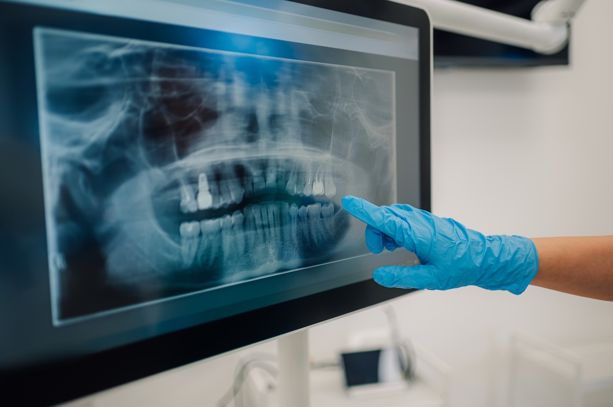
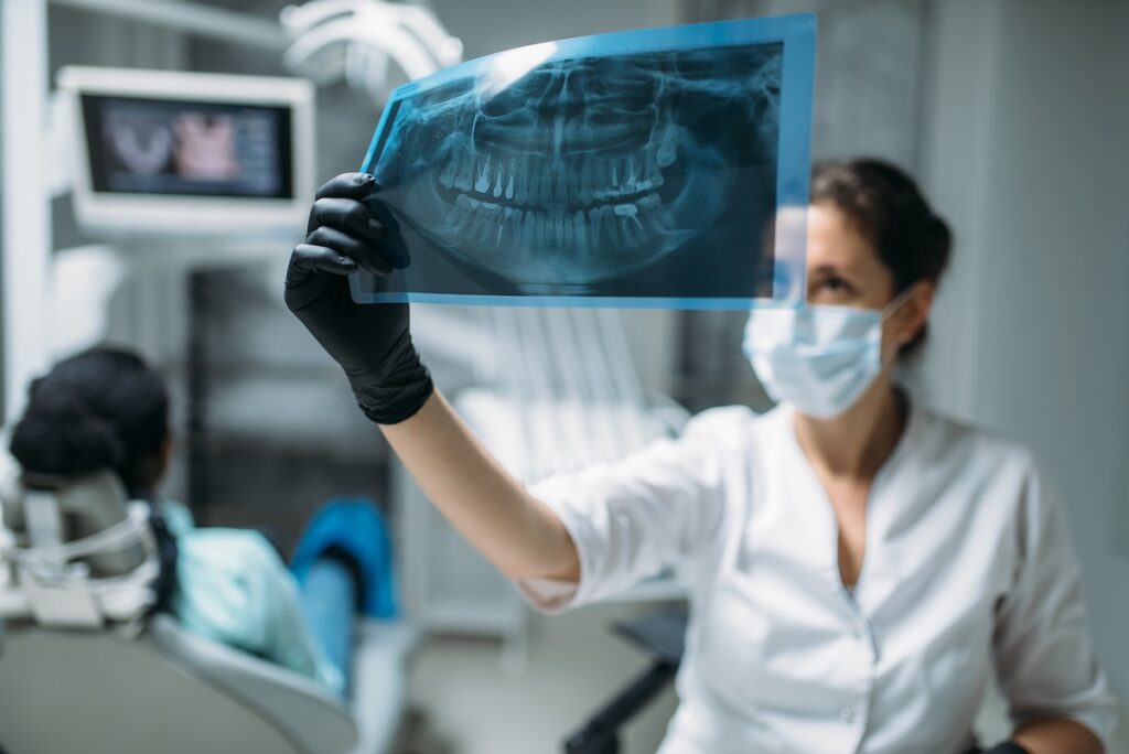
Panoramic X-ray (X-ray):
It shows the overall structure of the entire jaw and teeth. It is the first choice for detecting cavities and impacted teeth, and before orthodontic (braces) and implant procedures.
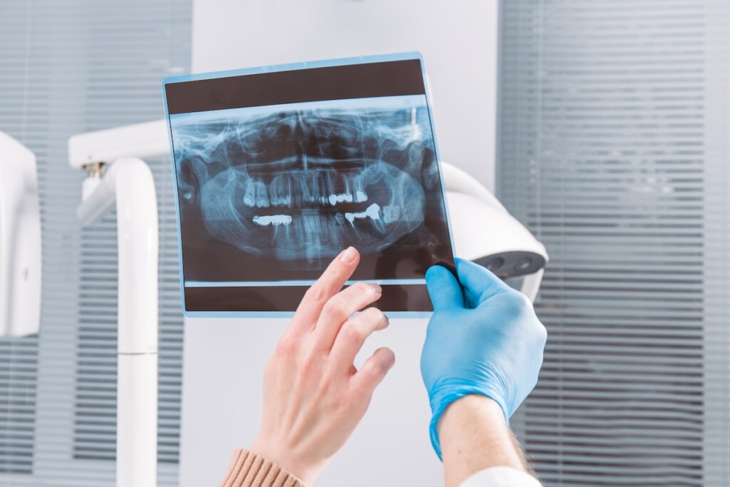
Reasons for Taking a Panoramic X-ray
• General Oral Health Check: A panoramic view of the jaw structure is taken during the first dental examination.
• Monitoring Tooth Development: In children, the eruption direction of permanent teeth, the condition of baby teeth, and jaw development are monitored.
• Treatment Planning: It is used for general evaluation before many procedures such as prosthetics, implants, and braces.
3D Tomography (3D):
Provides a more detailed and three-dimensional image of the jaw. The volume of the jawbone, nerve canals, and root positions are examined. In impacted tooth surgeries, it is evaluated whether the teeth are close to the bone, and for implant or cyst operations, positioning is analyzed.
Areas of Use
• Implant Treatment: Bone density and suitability for implants are evaluated.
• Root Canal Treatment: Determination of root canal structure
• Impacted Teeth: Position of the teeth and their relationship with surrounding tissues
• Jaw Joint (TMJ): Examination of joint structure
• Root Infections: Detection of inflammation or pathological conditions in tooth roots
• Cyst and Tumor Detection: Hidden infections, fractures, cracks, and joint problems in the jawbone that cannot be seen during examination are analyzed
• Orthodontics: Analysis of teeth and jaw structure
Panoramic X-ray in Children
In children over the age of 6, X-ray imaging plays a very important role in monitoring oral development and detecting cavities that cannot be seen with the naked eye. At younger ages, X-rays are taken only if necessary after an oral examination.
• Periapical X-ray: Generally used in endodontics (root canal treatment). It shows only a few teeth and their root tips in detail. It is used during and after treatment to monitor roots and for localized infected teeth.
• Digital Intraoral Scanning (3D Scanning): Commonly used in prosthetics and clear aligner treatments with high precision. It is a radiation-free method that creates digital models of teeth, replacing traditional impression materials.


